Microelectronics Component Specialists


Other Analytical Services


Metallographic Cross-sectioning


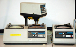
Metallographic cross-sectioning is the process of exposing the internal plane of interest of a sample for detailed examination. The process consists of mounting, sawing, grinding, polishing, and etching the sample so it can be analyzed using an optical microscope. A scanning electron microscope may also be used to verify and take a closer look at the microstructure that was observed under an optical microscope.


Back to Top


Optical Microscopy


An optical microscope is the most common (and the first to be invented) type of microscope.  It is used to view and inspect sample details that are too small to be viewed with the naked eye.  It is easy to use, and samples can be analyzed in air or water.  The images are in natural color with magnifications of up to 100 to 1000 times.  However, it does not have the same depth of focus and resolving power as the SEM


Back to Top


Particle Impact Noise Detection (PIND) Testing


PIND testing is used to detect loose particles inside a device cavity.  This test provides a means of identifying devices that contain particles with sufficient mass that upon impact with the case, excite the transducer.  It is a non-destructive test.


Applicable Specifications & Standards:

JEDEC JEP114, Guidelines for Particle Impact Noise Detection (PIND) Testing, Operator Training and Certification
MIL-STD-883 Method 2020, Particle Impact Noise Detection (PIND) Test
MIL-STD-750 Method 2052, PIND Test
MIL-STD-202 Method 217, Particle Impact Noise Detection (PIND)


Back to Top


Sputtering System


SIX SIGMA utilizes sputter-coater system to coat non-metallic samples with a thin layer of gold (other target materials include carbon, gold-palladium, platinum, and silver) to make them electrically conductive. This enables them to be viewed with a scanning electron microscope (SEM). This conductive coating inhibits charging, reduces thermal damage, and enhances secondary electron emission when the sample is examined under a SEM.


Back to Top